


Answer-First: Zombie cells are senescent skin cells that have stopped dividing but refuse to die. They release inflammatory signals (the SASP) that damage neighboring tissue, drive wrinkles and sagging, and slow wound healing. Cellular senescence is one of the 12 hallmarks of aging identified by López-Otín et al., Cell 2023 and accumulates rapidly in sun-exposed skin after age 40.
Last clinically reviewed by Dr. Dusan Sajic, MD, FRCPC, FAAD on 2026-05-29.
This article is part of our complete guide to the 12 Hallmarks of Aging. It is spoke #7 in the Pillar 1 series.
---
What are zombie cells?
Zombie cells are senescent cells: skin cells that should die but don't. They stop dividing yet remain metabolically active, releasing damaging signals into surrounding tissue. The term entered public science writing through Judith Campisi's lab, which first characterized the senescence-associated secretory phenotype in Coppé et al., PLOS Biology, 2008. López-Otín et al., Cell 2023 lists cellular senescence as one of 12 hallmarks of aging.
Why "zombie" stuck as the popular term
The metaphor works because senescent cells refuse to follow the normal cellular lifecycle. Healthy cells either divide or undergo apoptosis. Senescent cells do neither. They sit in the tissue, swollen and metabolically active, leaking inflammatory signals that turn nearby cells into more zombies.
In my dermatology practice, I think of zombie cells the way an electrician thinks of a corroded junction box. The wire still carries voltage, but the corrosion spreads. Removing the bad junction matters more than rewiring the whole circuit.
Why senescent cells accumulate in skin specifically
Skin is the body's most UV-exposed organ. Each unprotected exposure damages DNA, activates the p16INK4a and p21CIP1 cell-cycle arrest pathways, and converts a small fraction of fibroblasts into senescent cells. Over decades, the burden grows. Waaijer et al., Aging Cell 2012 measured p16INK4a-positive cells in human skin biopsies and found a steep age-related rise.
Citation capsule: Cellular senescence is one of the 12 hallmarks of aging identified by López-Otín et al., Cell, 2023. In skin, p16INK4a-positive senescent fibroblasts increase markedly with age, with Waaijer et al., Aging Cell, 2012 documenting roughly 6-fold higher senescent cell burden in older versus younger biopsies.
---
How do zombie cells form in skin?
Senescent cells form when a healthy cell hits a stress threshold and triggers permanent cell-cycle arrest. In skin, the dominant trigger is UV-induced DNA damage. Other inputs include oxidative stress, telomere attrition, and oncogene activation. The cell activates p16INK4a or p21CIP1, which lock the cell out of division (Hernandez-Segura et al., Trends in Cell Biology, 2018).
What turns a healthy cell into a zombie?
Four senescence triggers matter most for skin:
- UV-induced DNA damage. Even subclinical UV exposure activates the DNA damage response. In my TEDx talk on sunburns and zombie cells, I walked through how a single bad sunburn can produce a measurable senescent cell wave in the dermis within days.
- Oxidative stress. Mitochondrial dysfunction generates reactive oxygen species, damaging proteins, lipids, and DNA. Chen et al., Antioxidants, 2017 reviews ROS-driven senescence in skin.
- Telomere attrition. With each fibroblast division, telomeres shorten. Critical shortening triggers replicative senescence, the original mechanism described by Hayflick.
- Oncogene-induced senescence. A protective response that arrests pre-cancerous cells, but the resulting cells become inflammatory.
Why fibroblasts are the most common zombie cells
Dermal fibroblasts make collagen, elastin, and the extracellular matrix that gives skin its mechanical structure. They also live for decades. Long-lived, mechanically stressed, sun-exposed cells accumulate damage faster than short-lived cells like keratinocytes. Wang et al., Aging, 2020 shows fibroblasts as the dominant senescent population in photoaged skin.
Why senescence was originally protective
Senescence evolved as a tumor suppressor. A pre-cancerous cell that arrests cannot become a tumor. The system worked well for our ancestors who rarely lived past 40. Modern lifespans expose its dark side: senescent cells that accumulate over 60-80 years cause the inflammation and tissue dysfunction we see as aging.
Citation capsule: UV-induced DNA damage activates the p16INK4a and p21CIP1 cell-cycle arrest pathways, locking dermal fibroblasts into senescence. Hernandez-Segura et al., Trends in Cell Biology, 2018 reviews the molecular triggers. Wang et al., Aging, 2020 confirms fibroblasts as the dominant senescent population in photoaged human skin.
---

What do zombie cells do to your skin?
Zombie cells damage skin through the senescence-associated secretory phenotype, or SASP. Senescent fibroblasts leak inflammatory cytokines (IL-6, IL-8), matrix metalloproteinases (MMPs that chew up collagen), and chemokines that recruit immune cells. Coppé et al., PLOS Biology, 2008 catalogued the SASP secretome and showed it drives tissue dysfunction at low cell densities.
What is the SASP and why does it matter?
The SASP is the reason a small number of zombie cells causes outsized damage. Senescent fibroblasts make up only a few percent of total dermal cells in older skin, yet they secrete enough pro-inflammatory signal to alter tissue behavior at the millimeter scale.
The SASP cytokines most relevant to visible aging:
- IL-6 and IL-8: drive chronic low-grade inflammation
- MMP-1, MMP-3, MMP-9: degrade collagen and elastin, the structural proteins of skin
- TGF-β: disrupts wound healing
- CCL2 and CXCL10: recruit immune cells, amplifying inflammation
How one zombie cell turns its neighbors into zombies
The SASP is contagious at the tissue level. Acosta et al., Nature Cell Biology, 2013 showed that secreted factors from senescent cells induce paracrine senescence in neighboring healthy cells. One zombie cell creates a local pocket of inflammation; that pocket converts adjacent fibroblasts into more zombie cells. The cascade is why senescent cell burden grows non-linearly in older skin.
What you actually see in the mirror
The SASP cascade produces visible signs:
- Wrinkles and sagging from MMP-driven collagen and elastin loss
- Uneven texture and dullness from disrupted basement membrane signaling
- Slow wound healing from TGF-β dysregulation
- Age spots and dyspigmentation from inflammation-driven melanocyte dysfunction
The connection to chronic inflammation and inflammaging (Pillar 1 spoke #11) is direct: the SASP is one of the major sources of inflammaging in skin. The connection to skin microbiome dysbiosis (spoke #12) is mediated by inflammation altering the surface ecosystem.
Citation capsule: The senescence-associated secretory phenotype, characterized by Coppé et al., PLOS Biology, 2008, releases IL-6, IL-8, MMPs, and chemokines that drive collagen breakdown and chronic inflammation. Acosta et al., Nature Cell Biology, 2013 demonstrated paracrine senescence: one zombie cell can convert its neighbors into more zombies through secreted factors alone.
---
Can you remove zombie cells from skin?
Yes, in principle, but proven topical senolytics in dermatology remain limited. Senolytics are compounds that selectively kill senescent cells. The best-characterized systemic senolytics, dasatinib + quercetin and fisetin, were validated by Zhu et al., Aging Cell, 2015 and have shown senescent cell reduction in human trials (Hickson et al., EBioMedicine, 2019).
What senolytics actually exist?
Three classes of senolytic intervention exist today:
- Systemic small molecules: dasatinib + quercetin combination, fisetin, navitoclax. Oral or IV. Mostly studied in age-related disease, not cosmetic dermatology.
- Naturally derived senolytic compounds: quercetin, fisetin, hydroxytyrosol, piperlongumine. Some are bioavailable topically.
- Senomorphics: compounds that quiet the SASP without killing the cell. Bakuchiol, niacinamide, and certain peptides fall here.
What works topically: an honest take
After 22+ years in clinical dermatology, I treat the topical senolytic field with cautious optimism. The category is real. The science is moving fast. But the gap between "this compound kills senescent cells in a dish" and "this serum reduces wrinkles by clearing zombie cells in your skin" is enormous, and most marketing claims jump that gap without evidence.
What the literature actually supports today:
- Hydroxytyrosol (Hidrox) has documented antioxidant and anti-inflammatory effects that reduce SASP markers in keratinocytes. See our hydroxytyrosol research summary for the full citation set. Hydroxytyrosol is a key actor in Rejuvenat.
- Fisetin is a flavonoid with senolytic activity in vivo when delivered systemically. Topical formulations are emerging but lack the clinical validation of oral fisetin.
- Bakuchiol has anti-inflammatory and retinol-like effects with measurable reduction in oxidative stress, supporting senescence prevention. My peer-reviewed clinical study (Sajic et al., 2021, PMID 33740839) documents bakuchiol's tolerability and anti-aging effects.
- Quercetin appears in some topical formulations as an antioxidant. The dermal bioavailability is the limiting factor.
The honest framing for consumers
Today's cosmeceuticals reduce SASP signaling and prevent new senescence rather than killing existing zombie cells outright. The best-validated topical strategy is: prevent UV-induced senescence, quiet existing inflammation, and support cell turnover so damaged cells are replaced.
Citation capsule: The first true senolytic combination, dasatinib + quercetin, was validated by Zhu et al., Aging Cell, 2015 and tested in humans by Hickson et al., EBioMedicine, 2019, who showed measurable reduction in senescent cell markers. Topical senolytics for skin remain an emerging area with hydroxytyrosol, fisetin, and bakuchiol as the most evidence-supported candidates.
---
What is Dr. Sajic's protocol for senescence prevention?
A practical 5-step dermatologist protocol targets senescence at multiple intervention points. The strategy is to (1) block the dominant trigger, UV, (2) quiet existing SASP inflammation, (3) replace damaged cells with healthy ones through controlled cell turnover, and (4) avoid pro-senescence behaviors. Visible improvement typically takes 8-12 weeks of consistent use.
Step 1: Daily UV protection
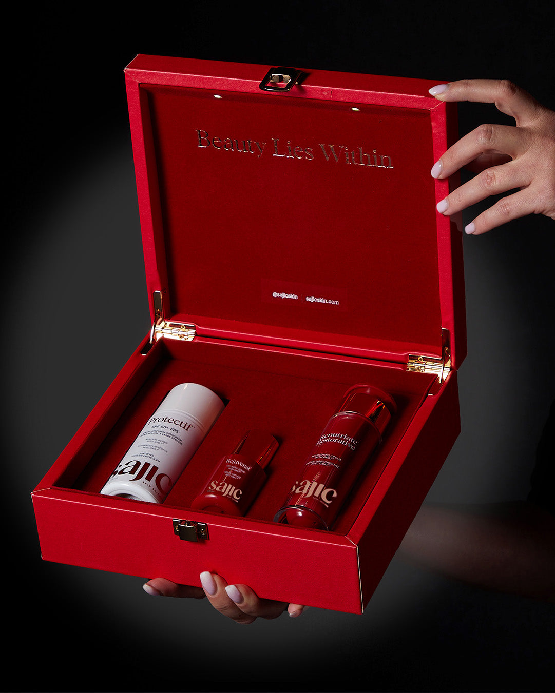
UV is the dominant senescence trigger in skin. Mineral sunscreen (zinc oxide-based) blocks UVA and UVB without the photoinstability of some chemical filters. See Protectif for the formulation I use in my own clinic.
Step 2: Antioxidant defense
Hydroxytyrosol (Hidrox) reduces oxidative stress that drives senescence. Rejuvenat combines hydroxytyrosol with the GMA7 delivery technology so the active reaches the dermis where senescent fibroblasts live.
Step 3: Bakuchiol or retinol for cell turnover
Healthy cell turnover replaces damaged fibroblasts before they accumulate as zombie cells. Bakuchiol is the better-tolerated option for sensitive skin; retinol is stronger but less forgiving. My peer-reviewed bakuchiol study (PMID 33740839) documents the tolerability advantage.
Step 4: Anti-inflammatory ingredients
Niacinamide, centella asiatica, and panthenol quiet the inflammation that propagates SASP signaling. They do not kill zombie cells, but they reduce the bystander damage zombie cells cause.
Step 5: Avoid pro-senescence behaviors
Smoking, chronic UV exposure without protection, repeated mechanical trauma, and chronic sleep deprivation all accelerate cellular senescence. The protocol works only if you remove the inputs that create new zombie cells.
Realistic timeline for visible results
In my clinical experience, patients on this protocol see meaningful texture and tone improvement at 8-12 weeks, with continued improvement through month 6. Senescence is a multi-decade process. Expect the protocol to slow accumulation and quiet inflammation, not erase decades of damage in a month.
---
Watch: How sunburns and zombie cells affect your health
[VIDEO EMBED: TEDx talk by Dr. Dusan Sajic - "How Sunburns and Zombie Cells Affect Your Health" - TEDxGreenhouse Road. Source: existing journal article 593951981789. Transcript and full discussion available at /blogs/journal/how-sunburns-and-zombie-cells-affect-your-health.]
In this TEDx talk, I walk through the link between UV-induced cellular senescence in skin and chronic systemic inflammation. The talk is the lay-audience companion to this article. Watch it for the narrative thread; come back here for the citations.
---
Frequently asked questions
Are zombie cells the same as cancer cells?
No. Zombie cells are the opposite of cancer cells. Cancer cells divide uncontrollably; senescent cells refuse to divide at all. Senescence evolved as a tumor suppressor mechanism (Campisi & d'Adda di Fagagna, 2007). The problem is that long-lived senescent cells secrete inflammatory signals that, paradoxically, can promote nearby tumor growth.
Can senescent cells in skin be reversed?
Most senescent cells cannot be returned to a healthy dividing state. The cell cycle arrest is enforced by p16INK4a and p21CIP1 and is essentially permanent (Hernandez-Segura et al., 2018). What can change: clearing senescent cells with senolytics, or quieting their SASP secretion with senomorphic compounds like bakuchiol and hydroxytyrosol.
What is the best ingredient for cellular senescence?
No single ingredient is "best." The strongest evidence-based stack combines daily mineral sunscreen (prevents new senescence), hydroxytyrosol or other antioxidants (reduces oxidative stress), and bakuchiol or retinol (drives healthy cell turnover). Senolytic clinical evidence is strongest for systemic dasatinib + quercetin and fisetin (Hickson et al., 2019), not topical formulations.
Does sunscreen prevent zombie cells?
Yes, daily broad-spectrum sunscreen is the single most effective senescence-prevention intervention available. UV is the dominant trigger of cellular senescence in skin (Wang et al., 2020). Zinc oxide mineral sunscreens block both UVA and UVB consistently, addressing the trigger most strongly linked to dermal fibroblast senescence.
How are zombie cells measured in skin?
Researchers use markers including p16INK4a expression, p21CIP1 expression, senescence-associated β-galactosidase (SA-β-gal) staining, and SASP cytokine panels. Waaijer et al., 2012 used p16INK4a immunohistochemistry on skin biopsies. No commercial at-home test is currently validated.
Are there topical senolytics that work?
The category is emerging. Topical hydroxytyrosol, fisetin, and bakuchiol have evidence for reducing oxidative stress and SASP markers, but no topical formulation has published clinical data showing bulk senescent cell clearance comparable to systemic dasatinib + quercetin. Treat marketing claims that promise senescent cell removal from a serum with skepticism.
How fast can you reduce SASP inflammation?
In my clinical practice, patients using consistent UV protection plus a hydroxytyrosol- and bakuchiol-based protocol typically report visible texture and tone improvement at 8-12 weeks. Inflammation markers measured in research settings can shift within 2-4 weeks of intervention, but visible skin changes lag the molecular changes.
---
About the author
Dr. Dusan Sajic, MD, FRCPC, FAAD is a board-certified dermatologist with 22+ years of clinical practice. He earned his MD from McMaster University, holds FRCPC and FAAD certifications, authored peer-reviewed research in the Journal of Cosmetic Dermatology (PMID 33740839), invented the patented GMA7 delivery technology, and is Past President of CLASS (Canadian Laser and Aesthetic Specialists Society). Read his full founder profile for credentials and verification links.
Last clinically reviewed: 2026-05-29.
---
References & Further Reading
- López-Otín C, Blasco MA, Partridge L, Serrano M, Kroemer G. The Hallmarks of Aging. Cell. 2013;153(6):1194-1217. doi.org/10.1016/j.cell.2013.05.039
- López-Otín C, Blasco MA, Partridge L, Serrano M, Kroemer G. Hallmarks of aging: An expanding universe. Cell. 2023;186(2):243-278. doi.org/10.1016/j.cell.2022.11.001
- Campisi J. Aging, cellular senescence, and cancer. Annu Rev Physiol. 2013;75:685-705. doi.org/10.1146/annurev-physiol-030212-183653
- van Deursen JM. The role of senescent cells in ageing. Nature. 2014;509(7501):439-446. doi.org/10.1038/nature13193
- Childs BG, Gluscevic M, Baker DJ, et al. Senescent cells: an emerging target for diseases of ageing. Nat Rev Drug Discov. 2017;16(10):718-735. doi.org/10.1038/nrd.2017.116
- Demaria M, Ohtani N, Youssef SA, et al. An essential role for senescent cells in optimal wound healing through secretion of PDGF-AA. Dev Cell. 2014;31(6):722-733. doi.org/10.1016/j.devcel.2014.11.012
- Wang AS, Dreesen O. Biomarkers of Cellular Senescence and Skin Aging. Front Genet. 2018;9:247. doi.org/10.3389/fgene.2018.00247
- Victorelli S, Lagnado A, Halim J, et al. Senescent human melanocytes drive skin ageing via paracrine telomere dysfunction. EMBO J. 2019;38(23):e101982. doi.org/10.15252/embj.2019101982
- Coppé JP, Desprez PY, Krtolica A, Campisi J. The senescence-associated secretory phenotype: the dark side of tumor suppression. Annu Rev Pathol. 2010;5:99-118. doi.org/10.1146/annurev-pathol-121808-102144
- Baker DJ, Childs BG, Durik M, et al. Naturally occurring p16(Ink4a)-positive cells shorten healthy lifespan. Nature. 2016;530(7589):184-189. doi.org/10.1038/nature16932
- Xu M, Pirtskhalava T, Farr JN, et al. Senolytics improve physical function and increase lifespan in old age. Nat Med. 2018;24(8):1246-1256. doi.org/10.1038/s41591-018-0092-9
- Tigges J, Krutmann J, Fritsche E, et al. The hallmarks of fibroblast ageing. Mech Ageing Dev. 2014;138:26-44. doi.org/10.1016/j.mad.2014.03.004
This article is for informational purposes only and does not constitute medical advice. Always consult a qualified healthcare professional for diagnosis and treatment decisions.


