


Answer-First: Inflammaging is chronic, low-grade inflammation that builds in tissues over decades and accelerates aging at the cellular level. In skin, it manifests as persistent redness, slow healing, diffuse sensitivity, and accelerated wrinkles. The term was coined by Franceschi, Annals NY Acad Sci, 2000 and is now recognized as one of the 12 hallmarks of aging by López-Otín et al., Cell 2023.
Last clinically reviewed by Dr. Dusan Sajic, MD, FRCPC, FAAD on 2026-05-22.
This article is part of our complete guide to the 12 Hallmarks of Aging. It is spoke #11 in the Pillar 1 series.
---
What is inflammaging?
Inflammaging is the chronic, low-grade, sterile inflammation that accumulates in tissues over decades. The term is a portmanteau of inflammation and aging, coined in Franceschi, Annals NY Acad Sci, 2000. López-Otín et al., Cell 2023 added chronic inflammation to the hallmarks of aging in their 2023 update, formalizing what gerontologists had argued for two decades.
Why "chronic + low-grade" matters
Acute inflammation is a controlled emergency response. A splinter triggers redness, swelling, heat, and pain that resolve within days. Inflammaging is the opposite. It is sub-clinical, persistent, and never fully resolves. The immune system idles at low throttle for years, slowly damaging the tissues it normally repairs.
The distinction is operational, not academic. A patient with acute contact dermatitis recovers in two weeks. A patient with inflammaging has skin that is, on a Tuesday afternoon with no obvious trigger, 15 percent more reactive than it was a decade ago. That baseline shift is the disease.
Why the immune system shifts with age
Three age-related immune changes drive inflammaging. First, the innate immune system loses calibration and overresponds to minor stimuli. Second, the adaptive immune system contracts, a process called immunosenescence (Franceschi & Campisi, J Gerontol, 2014). Third, senescent cells accumulate and pump out inflammatory signals via the SASP. The combination produces sustained, sterile, low-level inflammation across multiple organs.
Citation capsule: Inflammaging, coined in Franceschi, 2000 and characterized in Franceschi & Campisi, J Gerontol, 2014, is chronic, sterile, low-grade systemic inflammation. It was added to the canonical hallmarks of aging by López-Otín et al., Cell, 2023 and is mechanistically distinct from acute, pathogen-driven inflammation in trigger, intensity, and duration.
How does inflammaging show up in skin?
Inflammaging in skin presents as a constellation of low-intensity symptoms that patients often describe as "my skin is just different now." The dominant signs are persistent baseline redness, diffuse sensitivity that resists single-trigger explanations, slowed wound healing, and progressive barrier weakness (Pilkington et al., J Invest Dermatol, 2021).
Persistent redness and flushing
Inflammaged skin runs hot. Capillary dilation, low-level mast cell activation, and SASP-driven cytokine signaling produce a baseline pink that sits underneath everything else. It is not rosacea, though it overlaps. Patients often describe a flushed look in the mirror that sunscreen and gentle cleansing cannot mute.
Diffuse sensitivity
In my clinic, the diagnostic pattern is the patient who reacts to "everything." They cannot tolerate the vitamin C they used five years ago. They flush after a glass of wine that never bothered them. They feel their skincare more than they used to. The trigger is not any single ingredient. The threshold for inflammation has dropped.
Slow wound healing
Inflammaging dysregulates wound healing through TGF-β signaling and MMP overexpression. Sgonc & Gruber, Gerontology, 2013 reviewed age-related delays in cutaneous repair and connected them to chronic inflammatory tone. Even minor irritation, a popped pimple, a cuticle nick, takes longer to close in inflammaged skin.
Loss of barrier integrity
The stratum corneum thins. Lipid synthesis declines. Trans-epidermal water loss rises. A weak barrier admits more environmental insult, which provokes more inflammation, which further weakens the barrier. This is the inflammaging feedback loop in skin.
Connection to rosacea and menopause-onset sensitivity
Recent work proposes that rosacea sits on a continuum with inflammaging rather than being a discrete disease. Estrogen withdrawal in perimenopause and menopause amplifies the inflammatory tone and is a common inflection point in clinic (Lephart, Antioxidants, 2018). Patients who never had sensitive skin often develop it in their early 50s.
Citation capsule: Pilkington et al., J Invest Dermatol, 2021 reviewed inflammaging-specific skin manifestations and identified persistent low-grade erythema, slowed wound healing, and barrier compromise as the dominant clinical signs. Lephart, Antioxidants, 2018 tied estrogen-decline-driven inflammation to the menopausal sensitivity inflection that many patients report.

What causes inflammaging in skin?
Five mechanisms drive inflammaging in skin, and most patients have all five contributing simultaneously. The dominant inputs are senescent cells via the SASP, microbiome dysbiosis, cumulative UV damage, glycation, and repeated barrier disruption from over-aggressive skincare (Pilkington et al., J Invest Dermatol, 2021; Kammeyer & Luiten, Ageing Res Rev, 2015).
Senescent cells and the SASP
Zombie cells are an upstream driver. The senescence-associated secretory phenotype, characterized by Coppé et al., PLOS Biology, 2008, pumps out IL-6, IL-8, MMPs, and chemokines that activate the NF-κB pathway in neighboring tissue. The connection between cellular senescence (spoke #7) and inflammaging is direct: SASP is one of the largest sources of inflammaging in older skin.
Microbiome dysbiosis
Skin hosts a microbial ecosystem. With age, that ecosystem shifts. Diversity drops, opportunistic species rise, and the surface signaling environment moves toward pro-inflammatory tone. The full mechanism is the subject of Dysbiosis (spoke #12), the next post in this Pillar 1 series. For now: a disrupted microbiome feeds inflammaging.
Cumulative UV damage
UV is the dominant exogenous trigger. Each unprotected exposure activates DNA damage responses, generates reactive oxygen species, and adds to the senescent cell pool. Kammeyer & Luiten, Ageing Res Rev, 2015 reviewed oxidation-driven skin aging and the inflammatory cascade that follows.
Advanced glycation end products
Sugars cross-link with proteins to form AGEs, which bind RAGE receptors and activate NF-κB (Gkogkolou & Böhm, Dermatoendocrinol, 2012). AGE accumulation is a slow, chronic process that runs in parallel with UV damage and contributes to baseline inflammatory tone.
Repeated barrier disruption
In my 22+ years of practice, the most common iatrogenic trigger I see is over-cleansing combined with high-concentration acid stacking. Patients build a routine of foaming cleanser, AHA toner, BHA serum, retinol, and a vitamin C the next morning. Each step provokes a small inflammatory response. Stack them daily and the result is a chronically inflamed barrier. This is pro-inflammaging at home, dressed up as anti-aging.
Citation capsule: Inflammaging in skin is driven by senescent cell SASP signaling (Coppé et al., PLOS Biology, 2008), oxidative damage from UV (Kammeyer & Luiten, Ageing Res Rev, 2015), AGE-RAGE activation of NF-κB (Gkogkolou & Böhm, Dermatoendocrinol, 2012), and cumulative barrier disruption that perpetuates the inflammatory feedback loop.
What ingredients reduce inflammaging?
Five ingredient classes have peer-reviewed evidence for anti-inflammaging activity. The strongest are niacinamide, centella asiatica, hydroxytyrosol, bakuchiol, and ceramides. Each acts at a different point in the inflammatory cascade, which is why combinations outperform any single active.
Niacinamide (4-5%)
Niacinamide, vitamin B3, suppresses NF-κB signaling, reduces IL-6 and IL-8 secretion, and supports ceramide synthesis. Bissett et al., Dermatol Surg, 2005 showed measurable reductions in redness, blotchiness, and fine wrinkles at 5% over 12 weeks. The 4 to 5 percent range is the dose-response sweet spot.
Centella asiatica and madecassoside
Centella asiatica triterpenes, especially madecassoside and asiaticoside, suppress inflammatory cytokines and accelerate barrier recovery (Bylka et al., Adv Clin Exp Med, 2014). The ingredient class is one of the most robustly anti-inflammatory plant-derived options in dermatology.
Hydroxytyrosol (Hidrox)
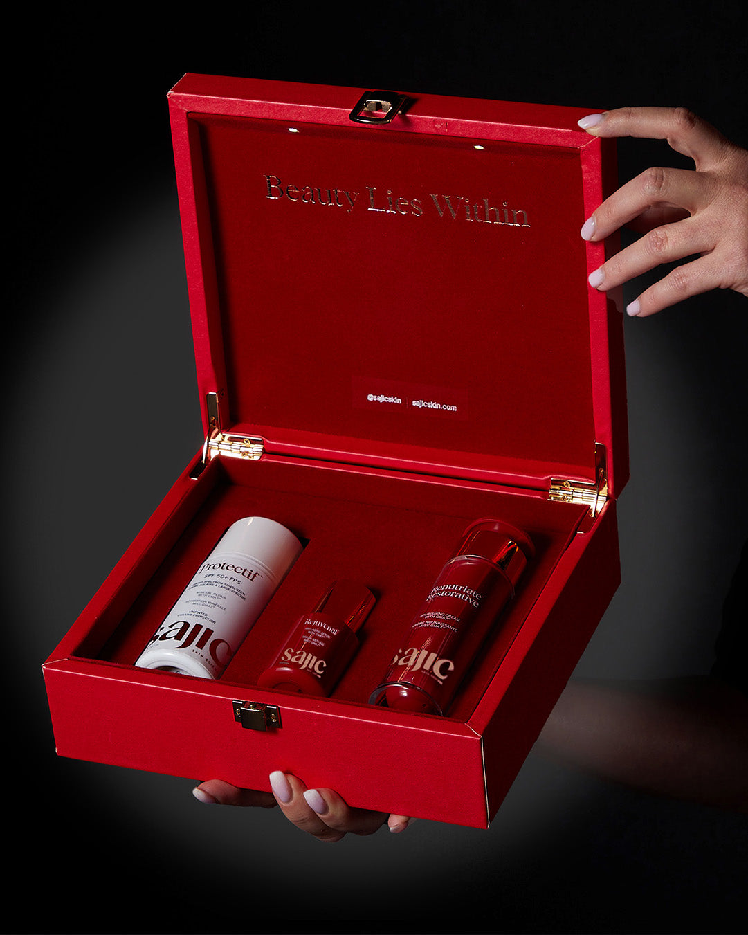
Hydroxytyrosol, the dominant polyphenol in olive fruit, is one of the most potent food-derived antioxidants known. It quenches reactive oxygen species, suppresses NF-κB activation, and reduces SASP markers in keratinocytes. Read our hydroxytyrosol research summary for the full citation set. Hydroxytyrosol is the lead actor in Rejuvenat, delivered into the dermis via GMA7 (Genoplex Microdelivery Activator).
Bakuchiol
Bakuchiol is anti-inflammatory in addition to its retinol-like effects on cell turnover. My peer-reviewed clinical study (Sajic et al., 2021, PMID 33740839) documented bakuchiol's tolerability in sensitive skin and measurable improvements in redness, fine lines, and barrier function over 12 weeks. It is one of the few actives that delivers anti-aging benefit without provoking the very inflammation we are trying to quiet.
Ceramides
Ceramide replenishment restores barrier integrity and breaks the inflammaging feedback loop. Spada et al., Clin Cosmet Investig Dermatol, 2018 demonstrated that ceramide-containing moisturizers measurably reduce TEWL and erythema in compromised skin. Renutriate is the formulation I use for barrier rebuild in inflammaged patients.
What to avoid
High-concentration AHA stacks, daily strong retinoids without recovery days, foaming cleansers with sulfates above 1 percent, and aggressive physical exfoliation all push the inflammatory cycle the wrong direction. Acute irritation, repeated, is pro-inflammaging.
What is Dr. Sajic's anti-inflammaging protocol?
A practical 5-step protocol targets inflammaging at multiple intervention points. The strategy stacks on top of, rather than replaces, the senescence prevention protocol from spoke #7. Visible reduction in baseline redness typically takes 8 to 12 weeks of consistent use.
Step 1: Daily UV protection
UV is the dominant exogenous driver. Mineral sunscreen with zinc oxide is the foundation. Without daily UV protection, every other step is fighting a leaking pipe.
Step 2: Antioxidant and anti-SASP defense
Hydroxytyrosol delivered into the dermis via GMA7 (Genoplex Microdelivery Activator) reduces oxidative stress and quiets SASP cytokine output. Rejuvenat is the formulation I prescribe at this step.
Step 3: Targeted anti-inflammatory actives
Niacinamide at 4 to 5 percent, centella asiatica, and panthenol applied morning and evening calm the inflammatory tone. These are senomorphics: they quiet the signal rather than killing the source cell.
Step 4: Barrier repair
Ceramides, cholesterol, and free fatty acids in the right ratio rebuild the stratum corneum. Renutriate targets this step. A repaired barrier admits less environmental insult, which lowers the inflammatory baseline.
Step 5: Eliminate pro-inflammaging behaviors
Smoking, chronic UV exposure, sleep deprivation, ultra-processed diets high in refined sugar, and over-aggressive skincare all push inflammaging upward. The protocol works only if you stop adding new fuel.
Realistic timeline
In my clinical experience, patients on this protocol see a measurable reduction in baseline erythema and sensitivity at 8 to 12 weeks, with continued improvement through month 6. Inflammaging took decades to build. Expect months, not weeks, to meaningfully shift it.
---
Frequently asked questions
What's the difference between inflammation and inflammaging?
Inflammation is the body's general response to injury or infection. It can be acute (sharp, time-limited) or chronic (sustained, often pathological). Inflammaging is a specific subtype: chronic, low-grade, sterile, age-associated inflammation that develops without an obvious trigger (Franceschi & Campisi, J Gerontol, 2014). The defining features are persistence, low intensity, and absence of pathogen.
Can inflammaging be reversed?
Inflammaging can be reduced, not erased. Targeting upstream drivers, senescent cells, microbiome dysbiosis, UV damage, and barrier dysfunction lowers baseline inflammatory tone over months. Research using senolytics, anti-SASP ingredients, and barrier repair shows measurable cytokine reductions in 8 to 12 weeks (Hickson et al., EBioMedicine, 2019; Bissett et al., Dermatol Surg, 2005). Reversal to a 25-year-old baseline is not realistic.
Is rosacea a form of inflammaging?
Rosacea is not officially classified as inflammaging, but the mechanisms overlap substantially. Both involve chronic low-grade dermal inflammation, vascular dysregulation, and innate immune over-activation. Pilkington et al., J Invest Dermatol, 2021 discusses the continuum. Some researchers now propose that adult-onset rosacea sits on the inflammaging spectrum rather than being a discrete disease.
Which is more important: anti-inflammaging or senolytics?
They target the same problem at different points. Senolytics aim to remove senescent cells. Anti-inflammaging aims to quiet the SASP signals those cells produce. In skin, the evidence base for senomorphic and anti-inflammaging ingredients is currently stronger than for topical senolytics, so the practical answer for cosmeceuticals is: prioritize anti-inflammaging now, watch the senolytics field as it matures.
Does diet affect skin inflammaging?
Yes. Dietary patterns high in refined sugar accelerate AGE formation (Gkogkolou & Böhm, Dermatoendocrinol, 2012), and ultra-processed diets are associated with elevated inflammatory markers. A Mediterranean-pattern diet rich in olive polyphenols, omega-3 fatty acids, and colorful plant antioxidants is the dietary side of an anti-inflammaging protocol. The skin is the largest organ; what you feed your immune system shows up there.
Are corticosteroids good for inflammaging?
No, not for chronic management. Topical corticosteroids quiet acute inflammation effectively, but long-term use thins the skin, weakens the barrier, and can rebound into worse inflammation when discontinued. They are pro-aging when used chronically. Anti-inflammaging is built on niacinamide, centella, hydroxytyrosol, bakuchiol, and ceramides, not steroid suppression.
How fast can I see results from anti-inflammaging skincare?
In my practice, patients typically report reduced flushing and improved tolerance to active ingredients at 6 to 8 weeks, with visible reduction in baseline redness and texture improvement at 12 weeks. Cytokine markers measured in research settings can shift in 2 to 4 weeks, but the visible skin change lags the molecular change.
---
About the author
Dr. Dusan Sajic, MD, FRCPC, FAAD is a board-certified dermatologist with 22+ years of clinical practice. He earned his MD from McMaster University, holds FRCPC and FAAD certifications, authored peer-reviewed research in the Journal of Cosmetic Dermatology (PMID 33740839), invented the patented GMA7 (Genoplex Microdelivery Activator) delivery technology, and is Past President of CLASS (Canadian Laser and Aesthetic Specialists Society). Read his full founder profile for credentials and verification links.
Last clinically reviewed: 2026-05-22.
---
References & Further Reading
- Franceschi C, Campisi J. Chronic inflammation (inflammaging) and its potential contribution to age-associated diseases. J Gerontol A Biol Sci Med Sci. 2014;69(Suppl 1):S4-S9. doi.org/10.1093/gerona/glu057
- Franceschi C, Garagnani P, Parini P, Giuliani C, Santoro A. Inflammaging: a new immune-metabolic viewpoint for age-related diseases. Nat Rev Endocrinol. 2018;14(10):576-590. doi.org/10.1038/s41574-018-0059-4
- Furman D, Campisi J, Verdin E, et al. Chronic inflammation in the etiology of disease across the life span. Nat Med. 2019;25(12):1822-1832. doi.org/10.1038/s41591-019-0675-0
- Pilkington SM, Bulfone-Paus S, Griffiths CEM, Watson REB. Inflammaging and the Skin. J Invest Dermatol. 2021;141(4S):1087-1095. doi.org/10.1016/j.jid.2020.11.006
- Zhuang Y, Lyga J. Inflammaging in skin and other tissues - the roles of complement system and macrophage. Inflamm Allergy Drug Targets. 2014;13(3):153-161. doi.org/10.2174/1871528113666140522112003
- López-Otín C, Blasco MA, Partridge L, Serrano M, Kroemer G. Hallmarks of aging: An expanding universe. Cell. 2023;186(2):243-278. doi.org/10.1016/j.cell.2022.11.001
- Bektas A, Schurman SH, Sen R, Ferrucci L. Aging, inflammation and the environment. Exp Gerontol. 2018;105:10-18. doi.org/10.1016/j.exger.2017.12.015
- Salminen A, Kaarniranta K, Kauppinen A. Inflammaging: disturbed interplay between autophagy and inflammasomes. Aging (Albany NY). 2012;4(3):166-175. doi.org/10.18632/aging.100444
- Pillai S, Oresajo C, Hayward J. Ultraviolet radiation and skin aging: roles of reactive oxygen species, inflammation and proteinases. Int J Cosmet Sci. 2005;27(1):17-34. doi.org/10.1111/j.1467-2494.2004.00241.x
- Fisher GJ, Kang S, Varani J, et al. Mechanisms of photoaging and chronological skin aging. Arch Dermatol. 2002;138(11):1462-1470. doi.org/10.1001/archderm.138.11.1462
- Calder PC, Bosco N, Bourdet-Sicard R, et al. Health relevance of the modification of low grade inflammation in ageing (inflammageing) and the role of nutrition. Ageing Res Rev. 2017;40:95-119. doi.org/10.1016/j.arr.2017.09.001
This article is for informational purposes only and does not constitute medical advice. Always consult a qualified healthcare professional for diagnosis and treatment decisions.


